


- 商品介绍
- 规格参数
- 包装参数
General description
Epidermal growth factor receptor (EC 2.7.10.1; UniProt P00533; also known as Proto-oncogene c-ErbB-1, Receptor tyrosine-protein kinase erbB-1) is encoded by the EGFR (also known as ERBB, ERBB1, HER1, NISBD2) gene (Gene ID 1956) in human. The epidermal growth factor receptor (EGFR; Proto-oncogene c-ErbB-1) is a ubiquitously receptor tyrosine kinase that is activated by a variety of ligands. Upon ligand-bound EGFR undergoes dimerization and autophosphorylation exposing cytoplasmic domains, which allows its interaction with adaptor proteins and induction of several signaling pathways including PI3K-AKT; PLC-gamma-PKC; Notch1; and Ras-Raf. Hence, it contributes to a diverse range of cellular processes, including cell proliferation. Several studies have reported that EGFR signaling plays an important role in the development of various tumors. EGFR is regulated by PTPRJ and PTPRK phosphatases, and by the endocytic machinery including EPS15 that mediates internalization and degradation of the receptor.
~170 kDa observed. Target band size appears larger than the calculated molecualar weight of 134.3 kDa due to glycosylation. Uncharacterized bands may be observed in some lysate(s).
Immunogen
GST-tagged recombinant human EGFR fragment correspoding to a cytoplasmic region C-terminal to the kinase domain.
Application
Detect EGFR using this mouse monoclonal , Cat. No. MABS835, validated for use in Flow Cytometry, Immunocytochemistry, and Western Blotting.
Flow Cytometry Analysis: 0.1 µg from a representative lot detected EGFR immunoreactivity in 4% paraformaldehyde-fixed, 0.2% Triton X-100-permeabilized A431 cells.
Immunocytochemistry Analysis: A 1:250 dilution from a representative lot detected plasma membrane EGFR immunoreactivity in 4% paraformaldehyde-fixed, 0.3% Triton X-100-permeabilized A431 cells.
Research Category
Signaling
Biochem/physiol Actions
Epitope region is present in spliced isoform 1 (p170), but not in isoform 2 (p60, Truncated, TEGFR), isoform 3 (p110) or isoform 4 reported by UniProt (P00533).
Physical form
Format: Purified
Purified mouse IgG2bκ in buffer containing 0.1 M Tris-Glycine (pH 7.4), 150 mM NaCl with 0.05% sodium azide.
Preparation Note
Stable for 1 year at 2-8°C from date of receipt.
Analysis Note
Evaluated by Western Blotting in A431 cell lysate.
Western Blotting Analysis: 0.1 µg/mL of this antibody detected EGFR in 10 µg of A431 cell lysate.
Other Notes
Concentration: Please refer to lot specific datasheet.
Replaces: 04-338
Disclaimer
Unless otherwise stated in our catalog or other company documentation accompanying the product(s), our products are intended for research use only and are not to be used for any other purpose, which includes but is not limited to, unauthorized commercial uses, in vitro diagnostic uses, ex vivo or in vivo therapeutic uses or any type of consumption or application to humans or animals.
| biological source | mouse |
| Quality Segment | 100 |
| conjugate | unconjugated |
| antibody form | purified immunoglobulin |
| antibody product type | primary antibodies |
| clone | 7F12.1, monoclonal |
| species reactivity | human |
| technique(s) | flow cytometry: suitable,immunocytochemistry: suitable,western blot: suitable |
| isotype | IgG2bκ |
| NCBI accession no. | NP_005219 |
| UniProt accession no. | P00533 |
| shipped in | ambient |
| target post-translational modification | unmodified |
| Gene Information | human ... EGFR(1956) |
| 长度(mm) | |
| 宽度(mm) | |
| 高度(mm) | |
| 重量(kg) |
-
询价
-
询价
-
询价
-
¥ 2068.25
-
¥ 3109.49
-
询价
-
询价
-
询价
-
询价
-
询价
-
询价
-
询价
-
询价
-
询价
-
¥ 5167.58
-
¥ 2883.61
-
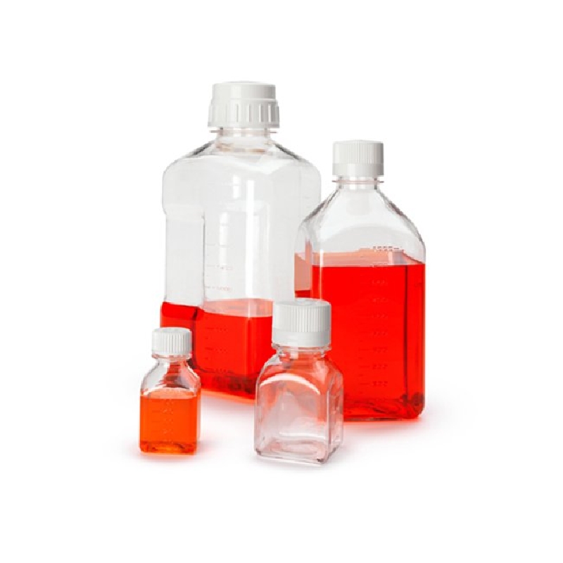
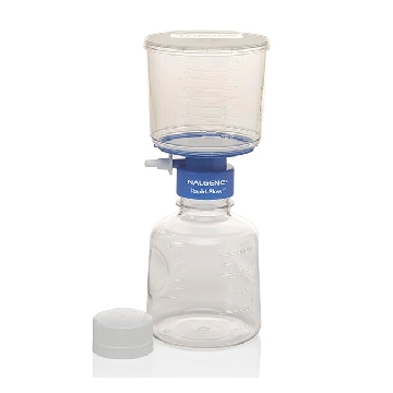
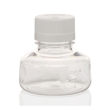
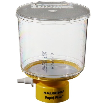 瓶顶过滤器-500ml容量,MF75TM系列,聚苯乙烯外壳,75mmSFCA滤膜,孔径0.2um,适合瓶颈尺寸45mm,12/箱,291-4520,Nalgene,Thermofisher,赛默飞世尔
¥ 2856.42
-

询价
-
¥ 2268.97
-
¥ 1749.15









 微信小程序
7X24小时在线咨询
微信小程序
7X24小时在线咨询