



- 商品介绍
- 规格参数
- 包装参数
General description
109.5 kDa calculated. ~150 kDa reported (Piela-Smith, T.H., and Korn, J.H. (1995). Cell Immunol. 1995 Apr 15 162(1):42-48) due to glycosylation.
Aminopeptidase N (EC 3.4.11.2; UniProt P15144; also known as Alanyl aminopeptidase, Aminopeptidase M, AP-M, AP-N, CD13, gp150, hAPN, Microsomal aminopeptidase, Myeloid plasma membrane glycoprotein CD13) is encoded by the ANPEP (also known as APN, CD13, PEPN) gene (Gene ID 290) in human. Aminopeptidase N (CD13) is a 150-kDa membrane glycoprotein belonging to the superfamily of zinc metalloproteases. CD13 preferentially cleaves N-terminus neutral amino acids, most notably alanine residue, and is widely expressed as a homodimer of 280 kDa on the cell surface in many tissues, including intestinal epithelia and the nervous system. CD13 is involved in many physiological processes such as antigen presention regulation, differentiation, proliferation, apoptosis, chemotaxis, phagocytosis, pain sensation, adhesion, viral infection, cancer metastasis and angiogenesis. CD13 mediates its biological functions through its role as a receptor or co-receptor in cellular signaling, as well as via peptidase activity. CD13 is a receptor for human coronavirus (HCoV) and cytomegalovirus (HCMV), and many proteins are found in complex with CD13, including galectin-3, galectin-4, Grb2, IgG receptors (Fc Rs), reversion-inducing cysteine-rich protein with kazal motifs, Sos, and the pro-inflammatory cytokine 14-3-3 . Human CD13 consists of a short N-terminal cytoplasmic end (a.a. 2-8), a transmembrane domain (a.a. 9-32), and a large extracellular portion (a.a. 33-967) composed of a Ser/Thr-rich region (a.a. 33-68) and the metalloprotease domain (a.a. 69-967), including an HCoV-229E-interacting region (a.a. 26-353) and a Substrate-binding region (a.a. 352-356).
Immunogen
Epitope: extracellular domain
Human foreskin fibroblasts.
Application
Electron Microscopy Analysis: A representative lot detected aminopeptidase N/CD13 on the surface of 4% paraformaldehyde-fixed human foreskin fibroblasts by EM (Piela-Smith, T.H., and Korn, J.H. (1995). Cell Immunol. 1995 Apr 15 162(1):42-48).
ELISA Analysis: A representative lot detected aminopeptidase N/CD13 on the surface of adherent human foreskin fibroblasts by "cell ELISA" (Piela-Smith, T.H., and Korn, J.H. (1995). Cell Immunol. 1995 Apr 15 162(1):42-48).
Flow Cytometry Analysis: Representative lots detected aminopeptidase N/CD13 on the surface of human foreskin fibroblasts, but not human squamous cell carcinoma cells A-431 and NCI-H292 (Piela-Smith, T.H., and Korn, J.H. (1995). Cell Immunol. 1995 Apr 15 162(1):42-48; Chen, L.L., et al. (1993). J. Tissue Cult. Methods.15:1-10).
Immunoprecipitation Analysis: A representative lot immunoprecipitated ~150 kDa aminopeptidase N/CD13 from human foreskin fibroblast (FB) lysate (Piela-Smith, T.H., and Korn, J.H. (1995). Cell Immunol. 1995 Apr 15 162(1):42-48).
Immunoaffinity Purification: A representative lot was conjugated to magnetic beads and employed to remove fibroblasts from primary cultures of squamous cell carcinoma of the head and neck (Chen, L.L., et al. (1993). J. Tissue Cult. Methods.15:1-10).
Research Category
Apoptosis & Cancer
This mouse monoclonal Anti-Aminopeptidase N/CD13 Antibody, clone BR2, Cat. No. MABF911 detects levels of Aminopeptidase N/CD13, and has been published and validated for use in Electron Microscopy, ELISA, Flow Cytometry, Immunoaffinity Purification, and Immunoprecipitation.
Biochem/physiol Actions
Clone BR2 targets aminopeptidase N/CD13 extracellular domain without inhibiting its enzymatic activity (Piela-Smith, T.H., and Korn, J.H. (1995). Cell Immunol. 1995 Apr 15 162(1):42-48). Clone BR2 specifically stains human foreskin fibroblasts, but not HUVECs, human intestinal SMCs, human keratinocytes (RHEK-1), human squamous cell carcinoma lines A-431 and NCI-H292, nor does it cross-react with rat or murine fibroblasts (Chen, L.L., et al. (1993). J. Tissue Cult. Methods.15:1-10).
Physical form
Format: Purified
Protein G purified.
Purified mouse monoclonal IgG1κ in buffer containing 0.1 M Tris-Glycine (pH 7.4) 150 mM NaCl with 0.05% sodium azide
Preparation Note
Stable for 1 year at 2-8°C from date of receipt.
Analysis Note
Evaluated by Flow Cytometry in human PBMCs.
Flow Cytometry Analysis: 0.1 µg of this antibody detected aminopeptidase N/CD13-positive granulocytes among human PBMCs.
Other Notes
Concentration: Please refer to lot specific datasheet.
Disclaimer
Unless otherwise stated in our catalog or other company documentation accompanying the product(s), our products are intended for research use only and are not to be used for any other purpose, which includes but is not limited to, unauthorized commercial uses, in vitro diagnostic uses, ex vivo or in vivo therapeutic uses or any type of consumption or application to humans or animals.
| biological source | mouse |
| Quality Segment | 100 |
| conjugate | unconjugated |
| antibody form | purified antibody |
| antibody product type | primary antibodies |
| clone | BR2, monoclonal |
| species reactivity | human |
| should not react with | mouse, rat |
| technique(s) | ELISA: suitable,affinity chromatography: suitable,electron microscopy: suitable,flow cytometry: suitable,immunoprecipitation (IP): suitable |
| isotype | IgG1κ |
| NCBI accession no. | NP_001141.2 |
| UniProt accession no. | P15144 |
| shipped in | ambient |
| target post-translational modification | unmodified |
| Gene Information | human ... ANPEP(290) |
| 长度(mm) | |
| 宽度(mm) | |
| 高度(mm) | |
| 重量(kg) |
-
询价
-
询价
-
¥ 1683.78
-
询价
-
询价
-
询价
-
询价
-

¥ 9737.84
-
询价
-
询价
-

询价
-
询价
-
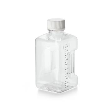
询价
-

询价
-
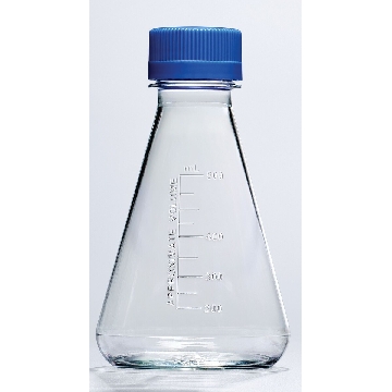
¥ 1509.00
-
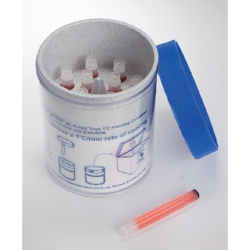
¥ 1586.69
-
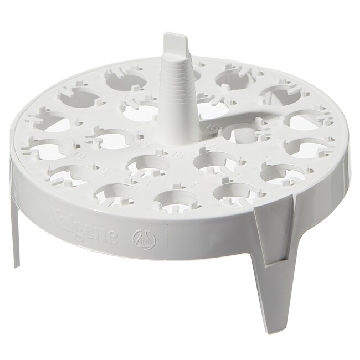
¥ 2099.27
-
¥ 3476.87
-
¥ 2754.43
-
¥ 56581.65









 微信小程序
7X24小时在线咨询
微信小程序
7X24小时在线咨询