





- 商品介绍
- 规格参数
- 包装参数
General description
The tumor suppressor p53 transcriptionally activates a number of genes including the WAF1/CIP1 gene in response to DNA damage. The ~21 kDa product of the WAF1 gene is found in a complex involving cyclins, CDKs, and PCNA in normal cells but not transformed cells and appears to be a universal inhibitor of CDK activity. One consequence of p21WAF1 binding to and inhibiting CDKs is the prevention of CDK-dependent phosphorylation and subsequent inactivation of the Rb protein which is essential for cell cycle progression. p21WAF1 is, therefore, a potent and reversible inhibitor of cell cycle progression at both the G1 and G2 checkpoints, presumably to allow sufficient time for DNA repair to be completed. Irreversible G1 or G2 arrest leads to apoptosis. While the role of p21WAF1 in apoptosis is less clear, it is known that p53-mediated apoptosis leads to increased WAF1 expression. Induction of p21WAF1 can occur by both p53-dependent and p53-independent mechanisms, in response to certain observed conditions. p21WAF1 has also been identified as a gene involved in cellular senescence, termed sdi1. Its overexpression was observed to inhibit cellular growth.
~21 kDa observed
Immunogen
Recombinant protein corresponding to human p21WAF1.
Application
Anti-p21WAF1 Antibody, clone EA10 is a Mouse Monoclonal Antibody for detection of p21WAF1 & has been validated in WB, FC.
Research Category
Epigenetics & Nuclear Function
Research Sub Category
Cell Cycle, DNA Replication & Repair
Western Blot Analysis: A representative lot from an independent laboratory detected p21WAF1 in non-irradiated and gamma irradiated CLL cells (Carter, A., et al. (2004). Br J Haematol. 127(4):425-428).
Immunohistochemistry Analysis: A representative lot from an independent laboratory detected p21WAF1 in normal human lower back tissue (el-Deiry, W. S., et al. (1995). Cancer Res. 55(13):2910-2919.).
Flow Cytometry Analysis: A representative lot from an independent laboratory detected p21WAF1 in non-irradiated and gamma irradiated CLL cells (Carter, A., et al. (2004). Br J Haematol. 127(4):425-428).
Physical form
Format: Purified
Protein G Purified
Purified mouse monoclonal IgG1κ supernatant in buffer containing 0.1 M Tris-Glycine (pH 7.4), 150 mM NaCl with 0.05% sodium azide.
Preparation Note
Stable for 1 year at 2-8°C from date of receipt.
Analysis Note
Control
HT-29 cell lysate
Evaluated by Western Blot in HT-29 cell lysate.
Western Blot Analysis: 1 µg/mL of this antibody detected p21WAF1 in 10 µg of HT-29 cell lysate.
Other Notes
Concentration: Please refer to the Certificate of Analysis for the lot-specific concentration.
Disclaimer
Unless otherwise stated in our catalog or other company documentation accompanying the product(s), our products are intended for research use only and are not to be used for any other purpose, which includes but is not limited to, unauthorized commercial uses, in vitro diagnostic uses, ex vivo or in vivo therapeutic uses or any type of consumption or application to humans or animals.
| biological source | mouse |
| Quality Segment | 100 |
| conjugate | unconjugated |
| antibody form | purified immunoglobulin |
| antibody product type | primary antibodies |
| clone | EA10, monoclonal |
| species reactivity | human |
| technique(s) | flow cytometry: suitable,western blot: suitable |
| isotype | IgG1κ |
| NCBI accession no. | NP_000380 |
| UniProt accession no. | P38936 |
| shipped in | wet ice |
| target post-translational modification | unmodified |
| Gene Information | human ... CDKN1A(1026) |
| 长度(mm) | |
| 宽度(mm) | |
| 高度(mm) | |
| 重量(kg) |
-
询价
-
¥ 1148.92
-
询价
-
¥ 1596.77
-
¥ 2019.57
-
询价
-
询价
-
询价
-

¥ 8135.09
-
询价
-
¥ 238.17
-
询价
-
询价
-
询价
-
¥ 6330.00
-
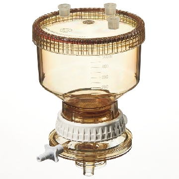
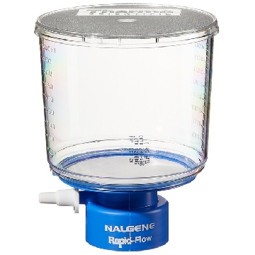 瓶顶过滤器-500ml容量,MF75TM系列,聚苯乙烯外壳,75mmPES滤膜,孔径0.45um,适合瓶颈尺寸33mm,12/箱,295-3345,Nalgene,Thermofisher,赛默飞世尔
¥ 1938.54
-
¥ 4897.21
-
¥ 745.60
-
¥ 5014.59
-
¥ 3031.24









 微信小程序
7X24小时在线咨询
微信小程序
7X24小时在线咨询