



- 商品介绍
- 规格参数
- 包装参数
General description
Stromal cell-derived factor 1 (SDF-1) is also known as C-X-C motif chemokine 12 (CXCL12), Intercrine reduced in hepatomas (IRH), and Pre-B cell growth-stimulating factor (PBSF). SDF-1 is a chemoattractant active on T-lymphocytes, monocytes, but not neutrophils. SDF-1 stimulates migration of monocytes and T-lymphocytes through its receptors, CXCR4 and CXCR7, and decreases monocyte adherence to surfaces coated with ICAM-1. SDF-1 plays a protective role after myocardial infarction, has critical functions during embryonic development and is required for B-cell lymphopoiesis, myelopoiesis in bone marrow and heart ventricular septum formation. Isoform Alpha and isoform Beta are ubiquitously expressed, with highest levels detected in liver, pancreas and spleen. Isoform Gamma is mainly expressed in heart. Isoform Delta, isoform Epsilon and isoform Theta have highest expression levels in pancreas.
~18 kDa observed. The calculated molecular weight of the SDF-1 monomer is 9 kDa However, the SDF-1 dimer has been observed at ~18 kDa (Veldkamp, C. T., et al. (2008). Sci Signal. 1(37):ra4.).
Immunogen
BSA-conjugated linear peptide corresponding to human SDF-1.
Epitope: N-terminus
Application
Detect SDF using this mouse monoclonal antibody, Anti-SDF-1 Antibody, clone K15C, Azide free validated for use in western blotting, IHC, Neutralizing & ELISA.
Research Category
Apoptosis & Cancer
Research Sub Category
Growth Factors & Receptors
Western Blotting Analysis: 0.375 µg/mL from a representative lot detected SDF-1 in 10 µg of HeLa and RAW264.7 cell lysates.
Western Blotting Analysis: A representative lot from an independent laboratory detected SDF-1alpha, SDF-1beta, and SDF-IP10 chimera, but not IP-10, IL-8, MDC, MIP-1b, eotaxin, and RANTES in WB (Coulomb-L′Hermin, A., et al. (1999). Proc Natl Acad Sci USA. 96(15):8585-8590.).
ELISA Analysis: A representative lot from an independent laboratory detected SDF-1alpha, SDF-1beta, and SDF-IP10 chimera, but not IP-10, IL-8, MDC, MIP-1b, eotaxin, and RANTES in ELISA (Coulomb-L′Hermin, A., et al. (1999). Proc Natl Acad Sci USA. 96(15):8585-8590.).
Immunohistochemistry Analysis: A representative lot from an independent laboratory detected SDF-1 in human fetal liver, lung, and kidney tissues (Coulomb-L′Hermin, A., et al. (1999). Proc Natl Acad Sci USA. 96(15):8585-8590.).
Neutralization Analysis: A representative lot from an independent laboratory attenuates the increased growth of MCF-7-ras tumor development in the presence of CAFs, decreases angiogenesis, and reduced recruitment of EPCs in tumors (Orimo, A., et al. (2005). Cell. 121(3):335-348.).
Immunhistochemistry Analysis: A representative lot from an independent laboratory detected SDF-1 in epidermal Dendritic Cells (DC) in normal and autoimmune diseased skin tissue (Pablos, J. L., et al. (1999). Am J Pathol. 155(5):1577-1586.).
Immunohistochemistry Analysis: A representative lot from an independent laboratory detected SDF-1 in mucosal surfaces of small intestine tissue, rectal tissue, vaginal tissue, and endocervical tissue (Agace, W. W., et al. (2000). Curr Biol. 10(6):325-328.).
Immunohistochemistry Analysis: A representative lot from an independent laboratory detected SDF-1 in mouse bone marrow sections (Dar, A., et al (2005). Nat Immunol. Epub 2005.).
Biochem/physiol Actions
This antibody recognizes the N-terminus of SDF-1. This antibody should detected SDF-1alpha and SDF-1beta.
Physical form
Format: Purified
Protein G Purified
Purified mouse monoclonal IgG2aκ in buffer containing PBS without preservatives.
Preparation Note
Stable for 1 year at -20°C from date of receipt.
Handling Recommendations: Upon receipt and prior to removing the cap, centrifuge the vial and gently mix the solution. Aliquot into microcentrifuge tubes and store at -20°C. Avoid repeated freeze/thaw cycles, which may damage IgG and affect product performance.
Analysis Note
Control
HEPG2 cell lysate
Evaluated by Western Blotting in HEPG2 cell lysate.
Western Blotting Analysis: 0.375 µg/mL of this antibody detected SDF-1 in 10 µg of HEPG2 cell lysate.
Other Notes
Concentration: Please refer to the Certificate of Analysis for the lot-specific concentration.
Disclaimer
Unless otherwise stated in our catalog or other company documentation accompanying the product(s), our products are intended for research use only and are not to be used for any other purpose, which includes but is not limited to, unauthorized commercial uses, in vitro diagnostic uses, ex vivo or in vivo therapeutic uses or any type of consumption or application to humans or animals.
| biological source | mouse |
| Quality Level | 100 |
| conjugate | unconjugated |
| antibody form | purified immunoglobulin |
| antibody product type | primary antibodies |
| clone | K15C, monoclonal |
| species reactivity | mouse, human |
| technique(s) | ELISA: suitable,immunohistochemistry: suitable,neutralization: suitable,western blot: suitable |
| isotype | IgG2aκ |
| NCBI accession no. | NP_000600.1 |
| UniProt accession no. | P48061 |
| shipped in | wet ice |
| target post-translational modification | unmodified |
| Gene Information | human ... CXCL12(6387) |
| 长度(mm) | |
| 宽度(mm) | |
| 高度(mm) | |
| 重量(kg) |
-

¥ 1563.36
-
询价
-
¥ 1866.29
-
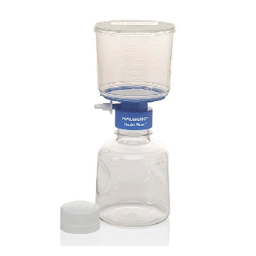
¥ 2068.25
-
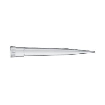
询价
-
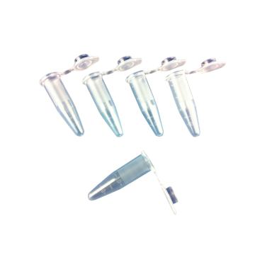
询价
-
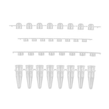
询价
-
询价
-
询价
-
询价
-
询价
-
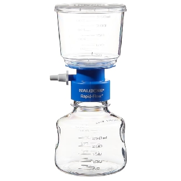
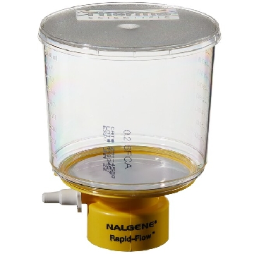 瓶顶过滤器-500ml容量,MF75TM系列,聚苯乙烯外壳,75mmSFCA滤膜,孔径0.2um,适合瓶颈尺寸45mm,12/箱,291-4520,Nalgene,Thermofisher,赛默飞世尔
¥ 2856.42
-
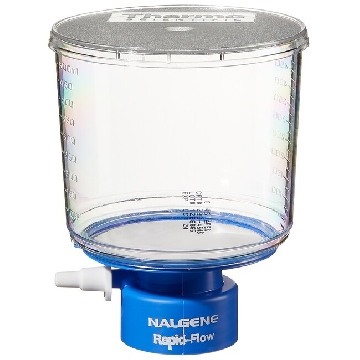 瓶顶过滤器-500ml容量,MF75TM系列,聚苯乙烯外壳,75mmPES滤膜,孔径0.45um,适合瓶颈尺寸33mm,12/箱,295-3345,Nalgene,Thermofisher,赛默飞世尔
¥ 1938.54
-
¥ 10277.12
-
¥ 3319.60
-
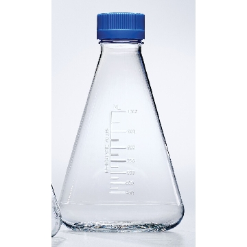
¥ 1564.00
-
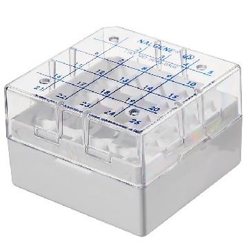
¥ 8137.12
-

¥ 954.17
-
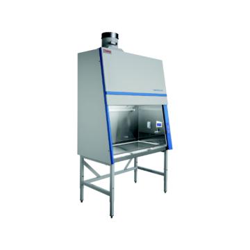
询价
-
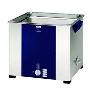
询价









 微信小程序
7X24小时在线咨询
微信小程序
7X24小时在线咨询