





- 商品介绍
- 规格参数
- 包装参数
General description
Myotubularin is a cytoplasmic phosphatase which belongs to the protein-tyrosine phosphatase family. It contains a GRAM domain and a myotubularin phosphatase domain. Myotubularin targets phosphoinositide substrates, including phosphatidylinositol 3-monophosphate (PI3P) and phosphatidylinositol 3,5-bisphosphate (PI(3,5)P2). It may also dephosphorylate phosphotyrosine- and phosphoserine-containing peptides. Myotubularin indirectly regulates many cellular processes that are facilitated by phosphoinositides, and plays an important role in the formation and morphology of membrane-bound intracellular structures such as vacuoles and mitochondria. It also regulates the transport of EGFR from late endosomes to lysosomes, resulting in the degradation of EGFR. Defects in myotubularin result in X-linked myotubular myopathy, a congenital muscle-wasting condition that affects the limb girdle, trunk, and neck muscles. It has also been implicated in other neuromuscular diseases, such as Charcot-Marie-Tooth disease type 4B.
~70 kDa observed
Immunogen
Epitope: N-terminus
Ovalbumin-conjugated recombinant protein corresponding to the N-terminus of human Myotubularin.
Application
Research Category
Signaling
Research Sub Category
Signaling Neuroscience
This Anti-Myotubularin Antibody, clone 1G6 is validated for use in western blotting, ICC & IP for the detection of Myotubularin.
Western Blotting Analysis: A 1:1,000 dilution from a representative lot detected Myotubularin in 10 µg of HL-60 cell lysate.
Western Blotting Analysis: A representative lot from an independent laboratory detected Myotubularin in lymphoblast lysates (Laporte, J., et al. (2002). J Cell Sci. 115(Pt 15):3105-3117.).
Immunoprecipitation Analysis: A representative lot from an independent laboratory immunoprecipitated Myotubularin from C2C12 cell lysates. (Laporte, J., et al. (2002). J Cell Sci. 115(Pt 15):3105-3117.).
Immunocytochemistry Analysis: A representative lot from an independent laboratory detected Myotubularin from C2C12 cells. (Buj-Bello, A. et al. (2002). Hum Mol Genet. 11(19):2297-2307.).
Physical form
Mouse monoclonal IgG1κ in ascites without preservatives.
Unpurified
Preparation Note
Stable for 1 year at -20°C from date of receipt.
Handling Recommendations: Upon receipt and prior to removing the cap, centrifuge the vial and gently mix the solution. Aliquot into microcentrifuge tubes and store at -20°C. Avoid repeated freeze/thaw cycles, which may damage IgG and affect product performance.
Analysis Note
Evaluated by Western Blotting in HeLa cell lysate.
Western Blotting Analysis: A 1:1,000 dilution of this antibody detected Myotubularin in 10 µg of HeLa cell lysate.
Disclaimer
Unless otherwise stated in our catalog or other company documentation accompanying the product(s), our products are intended for research use only and are not to be used for any other purpose, which includes but is not limited to, unauthorized commercial uses, in vitro diagnostic uses, ex vivo or in vivo therapeutic uses or any type of consumption or application to humans or animals.
| biological source | mouse |
| Quality Segment | 100 |
| conjugate | unconjugated |
| antibody form | ascites fluid |
| antibody product type | primary antibodies |
| clone | 1G6, monoclonal |
| species reactivity | mouse, yeast, human |
| technique(s) | immunocytochemistry: suitable,immunoprecipitation (IP): suitable,western blot: suitable |
| isotype | IgG1κ |
| NCBI accession no. | NP_000243 |
| UniProt accession no. | Q13496 |
| shipped in | wet ice |
| target post-translational modification | unmodified |
| Gene Information | human ... MTM1(4534) |
| 长度(mm) | |
| 宽度(mm) | |
| 高度(mm) | |
| 重量(kg) |
-
¥ 4173.75
-
询价
-
询价
-
询价
-
¥ 3442.25
-
询价
-
询价
-
询价
-
询价
-

询价
-
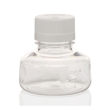
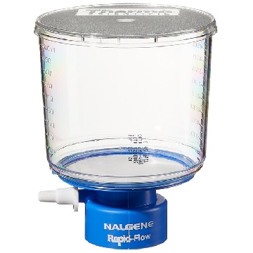 瓶顶过滤器-500ml容量,MF75TM系列,聚苯乙烯外壳,75mmPES滤膜,孔径0.45um,适合瓶颈尺寸33mm,12/箱,295-3345,Nalgene,Thermofisher,赛默飞世尔
¥ 1938.54
-
询价
-
¥ 2882.02
-
询价
-
¥ 1564.00
-
¥ 1749.15
-
¥ 787.34
-
¥ 2099.27
-
¥ 2911.61
-
¥ 9909.51









 微信小程序
7X24小时在线咨询
微信小程序
7X24小时在线咨询