



- 商品介绍
- 规格参数
- 包装参数
General description
140 kDa calculated
Collagen alpha-1(I) chain (UniProt P02452; also known as Alpha-1 type I collagen) is encoded by the COL1A1 gene (Gene ID 1277) in human. Collagen is the major component of the extracellular matrix (ECM) and forms the fibrils of tendons, ligaments, and bones. Type I collagen consists of two alpha I chains and one alpha 2 chain. Alpha-1 type I collagen is initially produced as a 1464-amino acid prepro-form with a signal peptide sequence (a.a. 1-22) and two propeptide sequences (a.a. 23-161 and a.a. 1219 –1464), the removal of which yields the mature alpha-1(I) chain. The mature alpha-1(I) chain is composed mostly of a large triple-helical region (a.a. 179-1192) sandwiched between two nonhelical segments known as the N-terminal telopeptide (a.a. 162-178; numbering based on the prepro-form) and the C-terminal telopeptide (a.a. 1193-1218; numbering based on the prepro-form). Collagen can be extracted from tissue via either enzymatic or non-enzymatic means. Collagen extracted using the proteolytic enzyme pepsin corresponds to the large triple-helical region, referred to as atelocollagen because both the N- and C-terminal telopeptides have been cleaved off by pepsin. On the other hand, collagen preparations obtained with non-enzymatic means (e.g. by acid extraction) have the intact telopeptides at both ends.
Immunogen
Epitope: C-terminal propeptide region
Human pro-collagen I.
Application
Immunohistochemistry Analysis: A 1:50 dilution from a representative lot detected Procollagen Type I in mouse skin, rat skin, and rat skeletal muscle tissue.
Immunocytochemistry Analysis: A representative lot detected type I procollagen immunoreactivity in human semitendinosus and gracilis tendon fibroblasts from patients undergoing reconstruction surgery after anterior cruciate ligament (ACL) rupture by fluorescent immunocytochemistry (Bayer, M.L., et al. (2012). Mech Ageing Dev. 133(5):246-254).
Flow Cytometry Analysis: A representative lot detected PICP+/CD45+ fibrocytes in lung cells from bleomycin-treated mice (Yeager, M.E., et al. (2012). Eur Respir J. 39(1):104-111).
Flow Cytometry Analysis: A representative lot detected higher numbers and percentages of circulating PICP+/CD45+ fibrocytes in peripheral blood samples from children/yound adults with pulmonary hypertension (PH) than in samples from healthy individuals (Reese, C., et al. (2014). Front Pharmacol. 5:141).
Immunohistochemistry Analysis: A representative lot detected cytoplasmic type I procollagen immunoreactivity in stromal cells from the lysed functionalis of frozen human menstrual endometria sections (Gaide Chevronnay, H.P., et al. (2009). Endocrinology. 150(11):5094-5105).
ELISA Analysis: A representative lot detected different age-dependency of procollagen type I C-terminal propeptide (PICP) immunoreactivity in the cruciate ligaments of osteoarthritis-/OA-prone Dunkin-Hartley (DH) guinea pigs and in age-matched Bristol strain 2 (BS2) control guinea pigs (Quasnichka, H.L., et al. (2005). Arthritis Rheum. 52(10):3100-3109).
Research Category
Cell Structure
Research Sub Category
Adhesion (CAMs)
This Anti-Procollagen Type I Antibody, CT, clone PCIDG10 (Ascites Free) is validated for use in Immunohistochemistry (Paraffin), Immunocytochemistry, Flow Cytometry and ELISA for the detection of Procollagen Type I.
Biochem/physiol Actions
Labels carboxy-terminal pro-peptide of collagen type I. Does not stain mature collagen fibers in tissue, but rather is localized intracellularly in cells producing pro-collagen I.
Physical form
Format: Purified
Protein G Purified
Purified mouse monoclonal IgG1κ antibody in buffer containing 0.1 M Tris-Glycine (pH 7.4), 150 mM NaCl without preservatives.
Preparation Note
Stable for 1 year at -20°C from date of receipt.
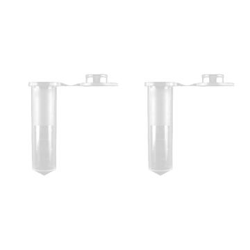
Handling Recommendations: Upon receipt and prior to removing the cap, centrifuge the vial and gently mix the solution. Aliquot into microcentrifuge tubes and store at -20°C. Avoid repeated freeze/thaw cycles, which may damage IgG and affect product performance.
Analysis Note
Evaluated by Immunohistochemistry in human bone tissue.
Immunohistochemistry Analysis: A 1:50 dilution of this antibody detected Procollagen Type I in human bone tissue.
Other Notes
Concentration: Please refer to lot specific datasheet.
Disclaimer
Unless otherwise stated in our catalog or other company documentation accompanying the product(s), our products are intended for research use only and are not to be used for any other purpose, which includes but is not limited to, unauthorized commercial uses, in vitro diagnostic uses, ex vivo or in vivo therapeutic uses or any type of consumption or application to humans or animals.
| biological source | mouse |
| Quality Segment | 100 |
| antibody form | purified immunoglobulin |
| antibody product type | primary antibodies |
| clone | PCIDG10, monoclonal |
| species reactivity | mouse, human, rat, guinea pig |
| species reactivity (predicted by homology) | bovine (based on 100% sequence homology) |
| technique(s) | ELISA: suitable,flow cytometry: suitable,immunocytochemistry: suitable,immunohistochemistry: suitable (paraffin) |
| isotype | IgG1κ |
| NCBI accession no. | NP_000079 |
| UniProt accession no. | P02452 |
| shipped in | dry ice |
| target post-translational modification | unmodified |
| Gene Information | human ... COL1A1(1277) |
| 长度(mm) | |
| 宽度(mm) | |
| 高度(mm) | |
| 重量(kg) |
-
询价
-
询价
-
¥ 2528.88
-
询价
-
¥ 1596.77
-
询价
-
询价
-
询价
-
询价
-
询价
-
询价
-
询价
-
¥ 3442.25
-
询价
-
询价
-
询价
-
¥ 3812.28
-
¥ 2268.97
-
询价
-
¥ 1564.00









 微信小程序
7X24小时在线咨询
微信小程序
7X24小时在线咨询