



- 商品介绍
- 规格参数
- 包装参数
General description
Proteoglycans (PG), present in all tissues, are one of the best-studied classes of matrix molecules. Over 50 members of PG have been characterized and studied for their biological significance. PG consist of a protein core to which glycosaminoglycan chains are covalently linked. Glycosaminoglycans (GAGs) are natural heteropolysaccharides that are present in every mammalian tissue. They are composed of repeating disaccharide units that consist of either sulfated or non-sulfated monosaccharides. Their molecular size and the sulfation type vary based on tissue type. The major types of GAGs found in mammalian tissues are: hyaluronan, chondroitin sulfate, and dermatan sulfate, heparin and heparan sulfate, and keratan sulfate. GAG chains are covalently bound to serine residues of the PG protein core via a tetrasaccharide linkage, consisting of xylose, two galactose residues and glucuronic acid. This antibody, clone PG-4, recognizes antigen that is associated with extracellular matrices and surfaces of cultured human dermal fibroblasts and the surfaces of cultured human adult keratinocytes. This antibody recognize native epitopes located within glycosaminoglycan chains and is detects both chondroitin sulfate (Versican) and dermatan sulfate (Decorin and Biglycan) proteoglycans. Treatment of PG with chondroitinases is reported to abolish its immunoreactivity for all antigens. (Ref.: Afratis, N et al. (2012). FEBS Journal 279 (7); 1177-1197; Sorrell, JM et al. (1999). Histochem. J. 31 (8); 549-558).
~60 kDa observed. Uncharacterized bands may be observed in some lysate(s).
Immunogen
Crude membrane fractions from adult human dermal fibroblasts from breast skin.
Application
Anti-Glycosaminoglycan, skin specific, clone PG-4, Cat. No. MABT819, is a mouse monoclonal antibody that detects glycosaminoglycan and has been tested for use in ELISA, Immunohistochemistry (Paraffin), and Western Blotting.
Research Category
Cell Structure
Western Blotting Analysis: A representative lot detected Glycosaminoglycan in Western Blotting applications (Sorrell, J.M., et. al. (1999). Histochem J. 31(8):549-58).
ELISA Analysis: A representative lot detected Glycosaminoglycan in ELISA applications (Sorrell, J.M., et. al. (1999). Histochem J. 31(8):549-58).
Immunohistochemistry Analysis: A representative lot detected Glycosaminoglycan in Immunohistochemistry applications (Sorrell, J.M., et. al. (1999). Histochem J. 31(8):549-58).
Biochem/physiol Actions
Clone PG4 detects glycosaminoglycan in skin cells.
Physical form
Format: Purified
Protein G purified
Purified mouse monoclonal antibody IgM in buffer containing 0.1 M Tris-Glycine (pH 7.4), 150 mM NaCl with 0.05% sodium azide.
Preparation Note
Stable for 1 year at 2-8°C from date of receipt.
Analysis Note
Evaluated by Immunohistochemistry (Paraffin) in human skin tissue.
Immunohistochemistry Analysis: A 1:250 dilution of this antibody detected Glycosaminoglycan in human skin tissue.
Other Notes
Concentration: Please refer to lot specific datasheet.
Disclaimer
Unless otherwise stated in our catalog or other company documentation accompanying the product(s), our products are intended for research use only and are not to be used for any other purpose, which includes but is not limited to, unauthorized commercial uses, in vitro diagnostic uses, ex vivo or in vivo therapeutic uses or any type of consumption or application to humans or animals.
| biological source | mouse |
| Quality Level | 100 |
| antibody form | purified immunoglobulin |
| antibody product type | primary antibodies |
| clone | PG-4, monoclonal |
| species reactivity | human, chicken, shark |
| packaging | antibody small pack of 25 μL |
| technique(s) | ELISA: suitable,immunohistochemistry: suitable (paraffin),western blot: suitable |
| isotype | IgMκ |
| shipped in | ambient |
| target post-translational modification | unmodified |
| Gene Information | human ... FAM20B(9917) |
| 长度(mm) | |
| 宽度(mm) | |
| 高度(mm) | |
| 重量(kg) |
-
询价
-
询价
-
¥ 4173.75
-
询价
-
询价
-
询价
-
询价
-
询价
-
询价
-
询价
-
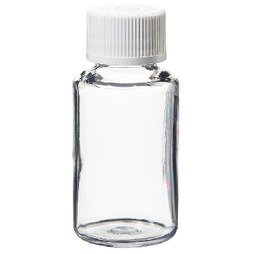
¥ 6330.00
-
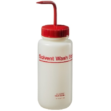
¥ 4390.38
-
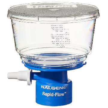
¥ 1653.62
-
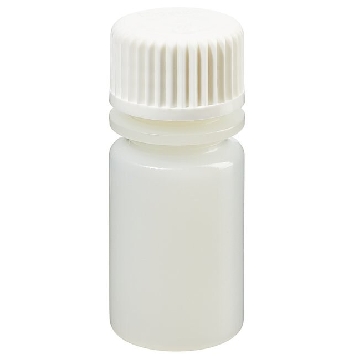
¥ 14407.09
-
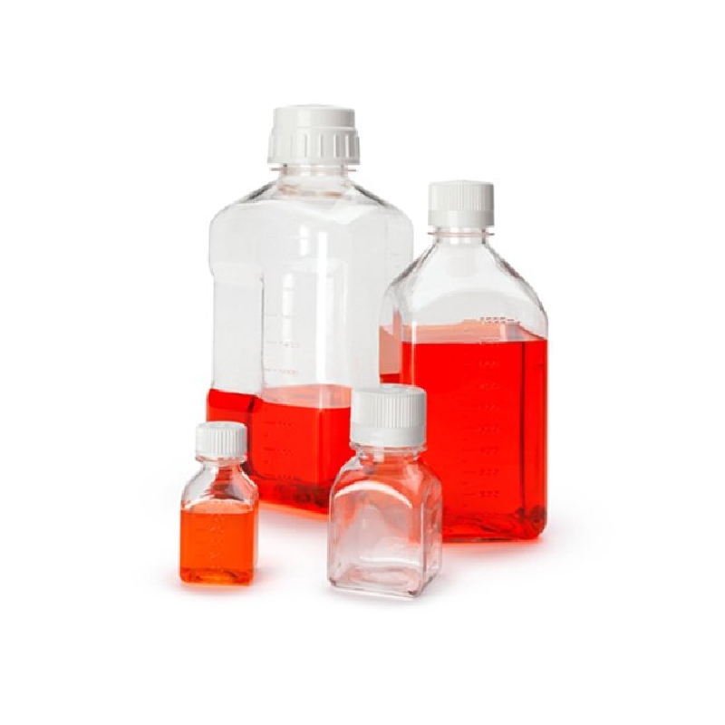
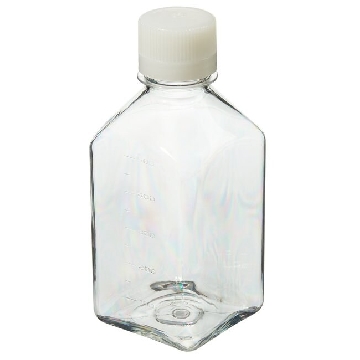 无菌,PETG 方形有刻度培养基瓶,带盖,聚对苯二酸乙二醇酯共聚物,天然高密度聚乙烯盖,500毫升容量,每箱40,40/箱,342020-0500,Nalgene,Thermofisher,赛默飞世尔
¥ 3684.34
-
¥ 2842.54
-
¥ 2666.57
-
¥ 2617.87
-
询价
-
¥ 1196.00









 微信小程序
7X24小时在线咨询
微信小程序
7X24小时在线咨询